Hemianopia: Types, causes, symptoms and treatment
What is hemianopia?
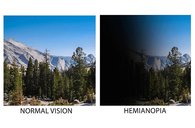
Hemianopia, also called hemianopsia, occurs when brain or optic nerve damage causes a person to lose sight in parts of their visual field. The condition affects sight out of each eye. The affected area in the field of vision may be the same or different out of each eye and depends on what part of the brain was damaged.
Hemianopia is usually seen in people after they’ve had a stroke or traumatic brain injury. Vision loss may be mild (dim or hazy) or severe (blindness). Depending on the cause, it’s possible for vision to improve with treatment, but some cases of hemianopia never recover.
Types of hemianopia
There are several types of hemianopia that are determined by the location and size of the visual field that is affected:
Homonymous – Most common type of hemianopia where the same half of each visual field is affected. The right half (right homonymous) or left half (left homonymous) of the visual field is lost, depending on which side of the brain is damaged. Damage will have occurred on the opposite side of the brain from where vision is lost. For example, injury on the right side of the brain will cause loss to the left side of the visual field.
Heteronymous – The opposite side of each eye is affected. Meaning the outer peripheral halves (bitemporal), or the inner halves (binasal) of the visual field are lost.
Superior – Loss of vision in the upper part of each visual field.
Inferior – Loss of vision in the lower part of each visual field.
Quadrantanopia – Vision loss in one quadrant of the visual field of both eyes.
SEE RELATED: What is blurry vision?
Partial vs. complete hemianopia
The difference between partial hemianopia and complete hemianopia is how much of a patient’s visual field is affected.
Partial hemianopia means the patient has no visual stimulus in one quadrant of the visual field. Complete hemianopia describes having no visual stimulus in half of their visual field.
Determining partial or complete hemianopia is part of the National Institutes of Health Stroke Scale (NIHSS). This scale is the most popular method for measuring the severity of a stroke by rating a patient’s neurological delays.
The third category of the NIHSS is the visual field test, where an eye care provider will measure hemianopia by having the patient look at them while covering one of their eyes. The eye care provider will hold up a certain number of fingers in each quadrant of the visual field and ask the patient to tell them how many fingers they’re holding up.
Once complete, the eye care provider will have the patient cover their other eye and perform the same test.
If the patient is able to see all the numbers flashed by the eye care provider, they will be ranked zero (0) on the NIHSS for “no vision loss.” Patients that have vision loss in one quadrant of their visual field for each eye will be ranked one (1) for “partial hemianopia.”
Vision loss in half of the visual field is ranked two (2) for “complete hemianopia.” Patients with bilateral blindness — the blindness being from any cause — are ranked three (3).
Hemianopia causes
The most common cause of hemianopia is stroke. However, brain tumors, inflammation and traumatic brain injury are other possible causes. Any damage to the optic nerves can also result in hemianopia.
Some less common conditions that have been known to cause hemianopia include:
Multiple sclerosis
Epilepsy
Neurosyphilis
Alzheimer’s disease
Lymphoma
Abnormal formation of blood vessels
Neurosurgical procedures
SEE RELATED: Causes of sudden and temporary double vision
Symptoms of hemianopia
Losing part of your visual field isn’t as obvious as you might think. However, there are some symptoms that may reveal hemianopia, including:
Inability to see objects located in the affected area of the visual field, such as cars in other lanes while driving, or food on a specific area of a plate
Visual hallucinations that appear as lights and various shapes
Turning the head away from the side affected by hemianopia
Bumping into things on the affected side
Disorientation in crowded environments
Drifting away from the affected visual field when walking
Reading only part of a block of text because the other part is “missing”
Diagnosis
Though hemianopia is a problem with the brain and not the eyes, you will need an eye exam to diagnose the condition. Your eye care provider will have you perform a visual field test that notes the location and size of the affected areas of your visual field.
The exam also includes having the eye care professional examine the inside of your eyes. This allows them to check the health of the retinas, macula and optic discs.
Additionally, the eye care professional will check the internal pressure of your eyes to look for other possible causes of peripheral (side) vision loss, such as glaucoma.
Besides an eye exam, your eye care professional will want you to obtain a head MRI (magnetic resonance imaging) to assess the health of your brain.
Treatment and prognosis
Treatment for hemianopia depends on the cause. For example, if a brain tumor is what caused the hemianopia, the appropriate treatment may be surgery, chemotherapy and/or radiation.
Once the tumor has shrunk or been removed, vision may improve. It’s also possible for people who suffered a brain injury or stroke to gain some vision back over time.
However, the prognosis for hemianopia in other situations may be limited, and there are many cases that never resolve. In these cases, treatment focuses on creating strategies to help patients learn to read and navigate their environment.
Reading strategies:
Rest a hand at the edge of a page, so it’s clearer to see where the margin ends.
Use a ruler, bookmark or straight edge to guide the eyes to the next line of text.
Hold text at various angles that allow it to be seen in full.
Increase the size of small eye movements as words are read along the line of text.
Navigation strategies:
Take a moment to pause and observe new environments. Move your head to scan your surroundings and take note of where objects and people are located.
Rely on your good visual field by directing your eyes toward it when moving around.
Walk with a partner who can cover your blind side and guide you with their arm, if necessary.
Work on card games or picture or word puzzles to improve coordination and eye scanning at close distances. This works best when done in real life, not on the computer.
In social situations, adjust yourself during conversation so people fall within your clear field of vision. This is also beneficial when watching television or a live performance.
Other treatment options:
Prismatic correction that helps expand the visual field
Compensatory training that improves visual search ability
Vision restoration therapy to improve eyesight
FIND AN OPTICIAN: if you're concerned about your vision, visit an optician near you.
Homonymous hemianopsia. Cleveland Clinic. April 2021.
Visual field loss in children. New England College of Optometry. Accessed October 2021.
What you need to know about hemianopia. Nova Vision. Accessed October 2021.
Visual field defects after stroke: A practical guide for GPs. Australian Family Physician. July 2010.
National Institutes of Health Stroke Scale. Radiology Review Articles. Accessed October 2021.
NIH Stroke Scale. National Institutes of Health. Accessed October 2021
Homonymous hemianopia: challenges and solutions. Clinical Ophthalmology. September 2014.
Stroke-related eye conditions. RNIB. Accessed October 2021.
Hemianopsia. StatPearls [Internet]. August 2021.
Your doctor thinks you have homonymous hemianopia. North American Neuro-Ophthalmology Society. 2016.
Page published on Tuesday, 17 May 2022


