Wavefront technology in eye exams
How wavefront technology improves eye care
Wavefront testing, also called aberrometry, is performed during some eye exams to analyze how light moves through the eye. It helps eye doctors detect and measure imperfections, or “aberrations,” in your vision. These are caused by irregularities in the eye’s structure that affect how you see.
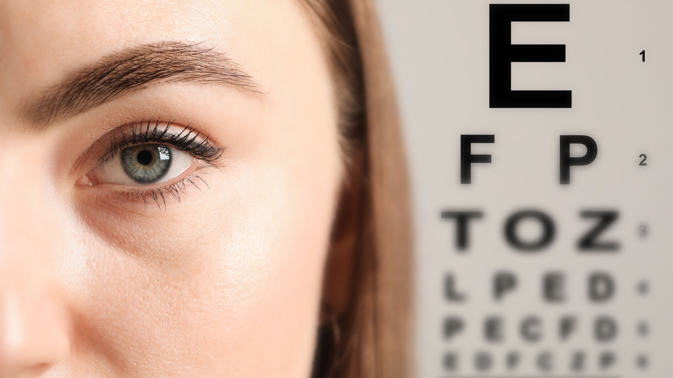
Wavefront, a technology originally developed for astronomy and later adapted for custom LASIK surgery, is used by some eye doctors to more precisely diagnose vision problems during eye exams. This type of testing could perhaps one day make the familiar eye chart obsolete.
What is a wavefront eye exam?
Most people have had eye exams with a device called a phoropter, which contains many lenses of different powers. An eye doctor changes the lenses in front of your eyes, asking which lens provides the sharpest image.
With this conventional approach, the information you give the eye doctor is very subjective and is based more on what you think you see instead of what you actually see.
Wavefront testing in eye exams works differently. Instead of relying on your feedback, a wavefront measurement is objective because vision errors can be identified automatically by the way light waves travel through the eye.
Someday, these detailed wavefront measurements may replace conventional eyeglass or contact lens prescriptions, which describe vision problems only in terms of the eye’s myopia (nearsightedness), hyperopia (farsightedness) and astigmatism.
Just as custom (or “wavefront-guided”) LASIK has the potential for producing sharper vision than conventional LASIK, high-definition glasses and contact lenses made with this advanced technology may also produce better visual clarity than their conventional counterparts.
What exactly does “wavefront” mean?
Imagine light traveling as a bundle of rays. If you draw lines perpendicular to the tips of a bundle of light rays, you get what is called a wavefront map. In an eye with perfect vision, the wavefront is perfectly flat. But the wavefront of an imperfect eye is irregular.
The types of distortions this wavefront acquires as it travels through the eye are called aberrations. They provide valuable information about vision errors and how to correct them.
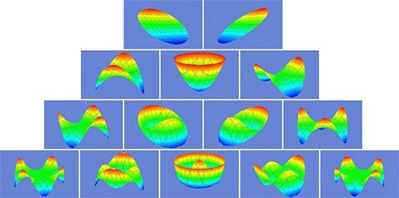
These are common shapes of aberrations created when a wavefront of light passes through eyes with imperfect vision.
What is wavefront technology (aberrometry)?
Aberrometry measures the way a wavefront of light passes through the cornea, crystalline lens and vitreous humor. These are the refractive (light-focusing) components of the eye. Distortions that occur as light travels through the eye are called aberrations, and each one represents a specific vision error.
Wavefront technology diagnoses both lower- and higher-order vision errors represented by the way the eye refracts or focuses light.
Lower-order aberrations are more common vision errors, such as nearsightedness, farsightedness and astigmatism. Higher-order aberrations are more complex vision errors, such as coma, trefoil and spherical aberration.
Wavefront and higher-order aberrations
Previously, with conventional methods of eye examinations, only lower-order vision errors could be diagnosed and treated. Higher-order aberrations were largely ignored by eye care professionals because their impact on vision was believed, at the time, to be slight and no feasible means existed to precisely identify or correct them.
Now that higher-order aberrations can be accurately defined by wavefront technology and corrected by new kinds of contact lenses, intraocular lenses and refractive surgery, they have become more important factors in eye exams.
Higher-order aberrations started receiving more attention because they were identified as sources of visual side effects after LASIK and other types of refractive surgery — causing halos, ghost images and other visual symptoms.
Newer wavefront-guided lasers used in vision correction surgery can reduce certain higher-order aberrations, which may improve visual performance, especially for low-light activities such as driving at night.
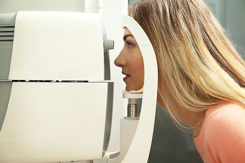
How is a wavefront measurement performed?
A wavefront diagnosis of the eye is performed with an aberrometer.
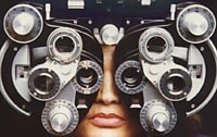
Traditional eye exams using phoropters with trial lenses can take a long time and rely on subjective feedback from patients. (Image: National Eye Institute)
Wavefront eye exams automatically and objectively measure vision errors in a few seconds and in much greater detail.
With most aberrometers, you place your chin on a chin rest. Next, you are asked to peer into the device and focus your eyes on a point of light. After a few seconds of silence (no feedback will be required of you), the wavefront measurement is completed and a wavefront map of your eye prints out for your eye care professional.
In general, an aberrometer uses a three-step process:
- Pupil measurement – Because a wavefront passes through the opening in the front of the eye (the pupil), your pupil diameter is measured. This measurement is used to derive a reference wavefront shape representing a theoretically perfect eye that has the exact same pupil size as yours.
- Light projection and reflection – A beam of light is projected into your eye and is reflected back by the retina. The wavefront of this reflected light is captured by the aberrometer. Because no eye is optically perfect, all such wavefronts will contain at least some distortions.
- Map creation – An aberration map of the eye is created by comparing the shape of the captured wavefront with that of the pre-programmed reference wavefront, measuring all points of difference between the two. Then, a wavefront map of your eye (sometimes called an “optical fingerprint” since no two eyes produce the exact same map) is created.
What does your eye’s wavefront map mean?
When interpreting the results of your eye’s wavefront map, keep in mind that the reference shape used for comparison is flat or two-dimensional.
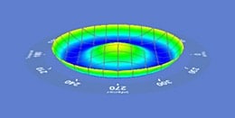
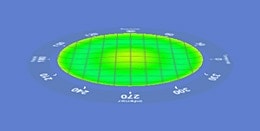
While no eye is ever perfect, a wavefront map of a theoretically perfect eye is a flat plane. Notice how this three-dimensional (3D) shape representing a vision error changes to almost a flat plane after the eye is corrected with wavefront-guided LASIK.
This flat, circular plane represents a theoretically perfect (emmetropic) eye that matches the diameter of your eye’s pupil exactly.
Your eye’s actual 3D wavefront map is created through comparison with this theoretically perfect, flat wavefront map. Three-dimensional distortions occur in the actual wavefront map based on how individual light rays are altered as they travel through the eye.
Imperfections in the cornea and lens can alter the path of light rays by causing them to speed up, slow down or bend (refract) incorrectly.
Other eye problems can also cause higher-order aberrations, including dry eye. Dry eye alters the eye’s tear film, which also helps refract light rays. Even cataracts (clouding of the eye’s natural crystalline lens) or scarring of the eye’s surface from eye surgery, disease or trauma can cause higher-order aberrations and focusing problems.
Based on the many different patterns that can occur in a wavefront, more than 60 aberrated shapes have been identified and classified as vision errors.
Wavefront measurements
Wavefront measurements that identify vision errors can be calculated in different ways. Currently, the method of choice is a mathematical expression called a Zernike polynomial.
Defined by Dutch physicist Fritz Zernike in 1934, this polynomial is well-suited to the task because it’s designed to meet the requirements of a circle.
Each Zernike polynomial describes a specific type of aberration across the entire wavefront of light after it passes through the eye. The sum of the Zernike polynomials equals a full description of all the aberrations or total refractive error in a given eye.
This finding is delivered in the form of a Zernike prescription and a topographic map — a detailed drawing of the shape of the aberrated wavefront. Together, they make up an aberration map or unique “optical fingerprint” that identifies the eye’s vision errors in detail.
How your wavefront map is used
The wavefront map is a complete, accurate description of all aberrations affecting your eye. Your eye doctor uses your wavefront map as a blueprint for custom-designing your vision correction, whether with:
- Refractive surgery (such as LASIK)
- Intraocular lenses (during cataract surgery)
- Contact lenses
- Glasses
Remember that you can never achieve a perfect wavefront because no eye is perfect. Even if you are diagnosed with a certain number of higher-order aberrations, this is not necessarily cause for concern, unless the degree of aberration causes significant vision problems, such as difficulty seeing at night.
Applications of wavefront technology
Wavefront technology allows for highly customized vision correction by mapping both lower- and higher-order aberrations. This information guides the design of surgical procedures and custom lenses, leading to potentially more precise treatment outcomes.
LASIK
Wavefront measurements provide a detailed 3D map of your eye’s aberrations to help customize LASIK treatment. There are two main approaches:
- Wavefront-guided (WFG) LASIK – This type uses a customized wavefront map to conduct the LASIK procedure. It can correct myopia, hyperopia and astigmatism, along with higher-order aberrations that can affect visual clarity even after correction of refractive errors.
- Wavefront-optimized (WFO) LASIK – This type is based on standard eye measurements rather than a custom wavefront map. It helps maintain the cornea’s natural shape while correcting refractive errors. WFO lowers the risk of visual distortions, such as halos and glare, that can result from conventional laser eye surgeries.
Custom lenses
Wavefront mapping is also used to design custom eyeglasses, specialty contact lenses and intraocular lenses placed during cataract surgery:
- Glasses and contact lenses – This technology detects higher-order aberrations that standard lenses may not be able to correct. Addressing these imperfections with customized lenses allows for sharper, clearer vision.
- Intraocular lenses (IOLs) – Wavefront technology can help guide IOL selection (such as light-adjustable lenses) or design custom IOLs that correct aberrations. This helps improve image quality and visual outcomes after cataract surgery.
As wavefront technology continues to evolve, it shows promise for even more accurate and personalized vision correction in the future.
READ MORE: Ophthalmoscopy: What is it?