Cavernous sinus
What is the cavernous sinus?
The cavernous sinuses are located in the head, behind the eyes and below the temporal lobe of the brain. They are important drainage spaces for several veins in the brain and eyes. They also serve as pathways for motor and sensory nerves that connect the brain to the eyes and face.
Problems in the cavernous sinuses are rare. However, their location and complex anatomy mean that any problems that do occur can be serious. They are very closely linked to the overall function of the eyes. They are also a potential pathway for infection. When bacteria or other pathogens from the face enter the cavernous sinus, infection can spread to the brain.
Anatomy of the cavernous sinus
There are many types of sinuses throughout the body. The word sinus is most often associated with the nasal cavity. However, the cavernous sinuses are a different type of sinus. They are connected to blood vessels in the head.
The cavernous sinuses (CS) are “paired” sinuses. There is one CS on each side of the cranial cavity, connected in the middle by the intercavernous sinuses. Inside, they look like they are divided into little caverns, which is where they get their name.
These caverns are made by a weblike series of thin, fibrous walls called septa.
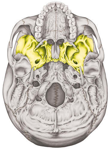
View of the sphenoid bone of the cranium.
The CS pair resides in the area of the skull called the central skull base. This area is partly made up of the sphenoid bone, which is shaped like butterfly wings. Each CS sits in one of its "wings." The pituitary gland sits in a depression in the center of the sphenoid bone.
These sinuses are a type of dural venous sinus. Dural means that they develop inside the dura mater. The dura mater is one of three layers of the protective tissue that wraps around the brain and spinal cord to keep them safe. Venous means that they act as drainage sites for blood carried through veins from the brain and face.
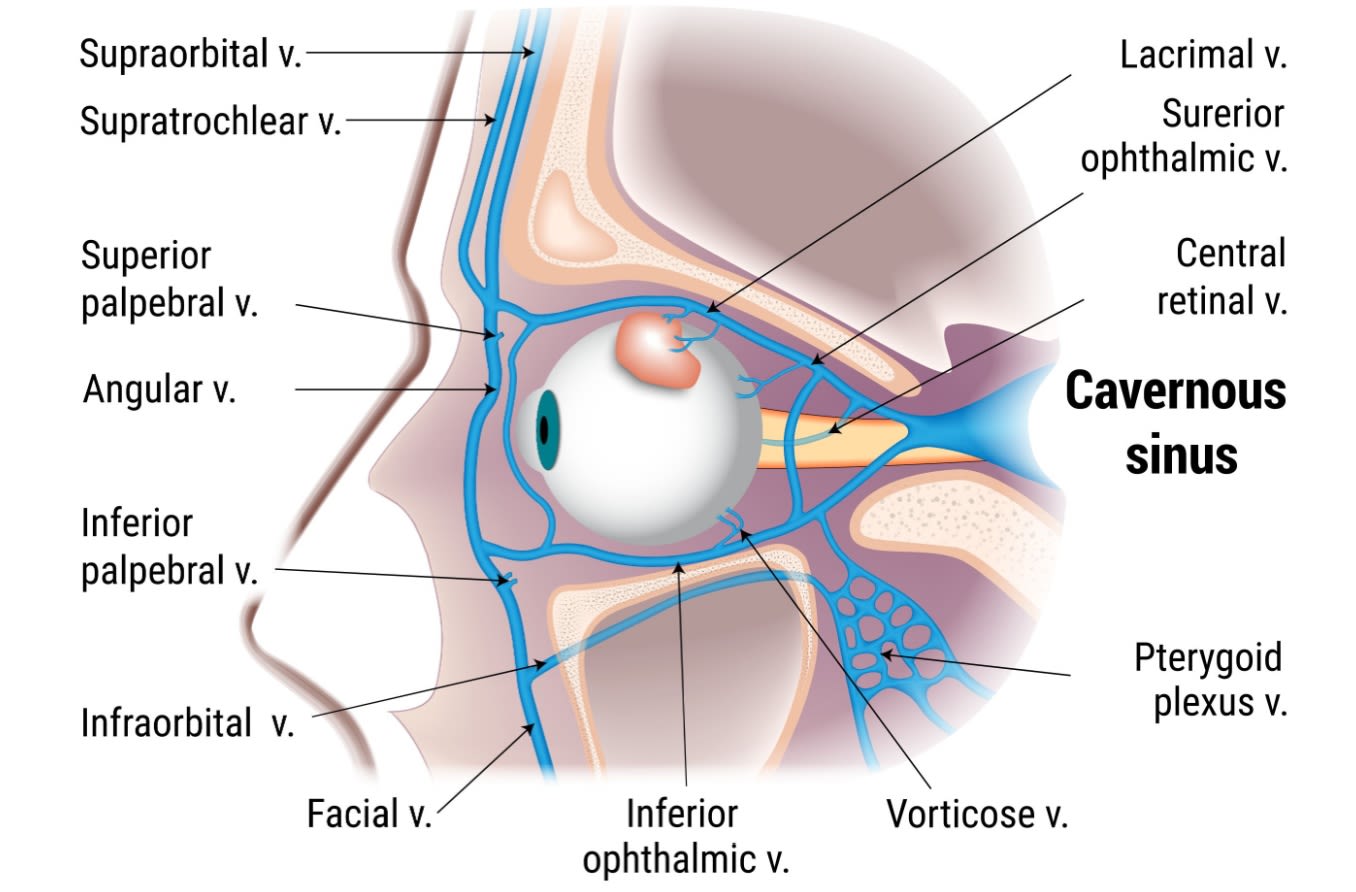
As part of the brain’s larger dural venous system, they connect to other dural sinuses. They also receive blood from veins, including the ophthalmic veins. The ophthalmic veins carry blood from the eyes and eye sockets (orbits), as well as from surrounding muscle and tissue and part of the nose. They join up to facial veins before draining into the cavernous sinuses.
The blood collected in the CS travels through the petrosal sinuses, then the sigmoid sinuses. The sigmoids transition into the internal jugular veins, which run through the neck on either side of the spine. These veins eventually merge with other veins to form the body’s largest vein: the superior vena cava.
Several sensory and motor nerves also thread through the CS. They use this pathway to reach the eyes and face. These important nerves include:
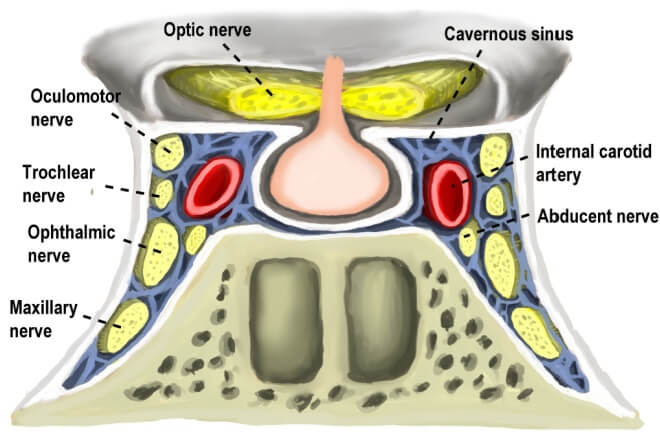
View of the cavernous sinus and the different nerves. Click image to enlarge.
Oculomotor nerve – A motor nerve that activates several muscles in and around the eyes. This includes muscles that lift the eyelid, rotate the eye up and down, and adjust the pupil and lens. CN III also controls coordination among these muscles during head or eye movement.
Trochlear nerve – A motor nerve that activates the muscles that rotate the eyes downward and to the side.
Ophthalmic nerve – The main sensory nerve for the eyes, forehead, scalp and parts of the nasal cavity. First of three branches of the trigeminal nerve.
Maxillary nerve – A sensory nerve related to the mid-face, the eyes' lacrimal glands and the nasal mucous glands. Second branch of the trigeminal nerve.
Abducens nerve – A motor nerve that activates the muscles that move the eyes toward the ear.
Segments of the internal carotid arteries (ICA) pass through each cavernous sinus, as well. They are among the most important arteries in the body.
Function of the cavernous sinus
The main function of the CS is to help drain deoxygenated venous blood from the brain, eyes and face. This is a vital step in the body’s circulatory system.
Their function as pathways for the ICA and cranial nerves is also significant. They are the most important dural sinuses because these vital structures pass through them. As a unit, the cavernous sinuses are referred to as the “anatomic jewel box.”
This "jewel box" can make it difficult to perform surgery in the CS when needed. Its location in the skull is also important. It can allow face or eye infections to spread to the cavernous sinus and then to the brain.
Conditions that can affect the cavernous sinus
Several conditions can impact the cavernous sinuses. Due to the sinuses' anatomy, these conditions can all cause similar symptoms. This set of symptoms is called cavernous sinus syndrome (CSS). It's a catch-all term to describe any disease that affects the nerves and/or arteries within the CS.
CSS can present with:
Proptosis/exophthalmos (bulging or protruding eye(s))
Chemosis (swelling of the conjunctiva and eyelids)
Ophthalmoplegia (eye muscle weakness or paralysis)
Horner’s syndrome (a combination of eyelid drooping, pupil constriction and an absence of facial sweating)
Trigeminal sensory loss (loss of feeling in the nerves that allow the face and eyes to sense pain, touch and temperature)
Any condition that can lead to CSS is potentially life-threatening and requires immediate medical attention. Although rare, tumors in and near the cavernous sinus are the most common cause of CSS. Trauma to the area and inflammation are also common causes.
Other conditions that can cause CSS include:
Cavernous sinus infection
The drainage connections between the facial vein, ophthalmic veins and cavernous sinuses create a pathway for infections to spread. If an infection spreads to the cavernous sinuses, it can then affect the cranial nerves there and cause serious complications.
It can also travel into the brain. Eye, nose, mid-face and mouth infections can potentially spread to the cavernous sinuses and then the brain. This cavernous sinus infection pathway is where the term danger triangle comes from. It is the reason behind the advice that you should not squeeze or pop pimples in this area.
These infections can also lead to cavernous sinus thrombosis.
Cavernous sinus thrombosis
Cavernous sinus thrombosis (CST) is very rare but dangerous. Thrombosis is when a blood clot (thrombus) in a vein or artery limits blood flow. The most common cause of CST is a bacterial infection. However, it can also be caused by:
Trauma to the CS
Protein deficiency
Oral contraceptives
Pregnancy
Immunosuppression
Blood clotting disorders
The most common symptoms of CST are fever and headache. If a blood clot becomes trapped in the cavernous sinus, it can also cause:
Edema (swelling) of the face and eye area
Ptosis (eyelid droop)
Proptosis/exophthalmos
Chemosis
Eye pain
Papilledema (swelling of the optic disc)
CST has high rates of serious complications (up to 40%) and even death (up to 20%). Complications can include eye muscle paralysis, cranial nerve palsy and blindness, among others. It requires IV antibiotics and immediate medical attention. CST is confirmed by a CT scan or an MRI.
Carotid-cavernous fistula
A fistula is an abnormal connection between two body parts. A carotid-cavernous fistula (CCF) is an irregular connection between the cavernous sinus and the carotid artery. A direct CFF results from a tear, usually from an injury. An indirect CCF is due to an underlying condition.
The fistula can cause blood flow to be rerouted. As a result, blood may not drain properly from the eye, causing it to bulge out.
Along with physical trauma, risk for CCF increases in women who are postmenopausal and in individuals with Ehlers-Danlos syndrome or fibromuscular dysplasia.
The most common symptom of a CFF is pulsatile exophthalmos (a pulsing, bulging eye). Other signs and symptoms may include:
Chemosis
Subconjunctival hemorrhage (blood in the white of the eye)
Proptosis/exophthalmos
Vision loss
Pulsatile tinnitus (a pulsing ringing in the ears)
Cranial nerve palsies (weakness or paralysis of the cranial nerves)
An indirect CFF may get better on its own. A direct CFF is typically treated by a neurosurgeon with various therapies or surgery.
When to see a doctor
Conditions that impact the cavernous sinuses are rare. However, they can lead to very serious complications, including blindness or even death. See your doctor right away if you experience any of the symptoms listed above.
It is also important to keep up with your yearly eye exams. Eye doctors can often see the signs of an issue before patients realize there is a cavernous sinus problem.
Shannon Belden also contributed to this article.
Imaging spectrum of cavernous sinus lesions with histopathologic correlation. RadioGraphics. April 2019.
Neuroanatomy, cavernous sinus. StatPearls [Internet]. July 2023.
Cavernous sinus syndrome. EyeWiki. American Academy of Ophthalmology. March 2024.
Management of cavernous sinus meningiomas: Consensus statement on behalf of the EANS skull base section. Brain and Spine. January 2022.
Surveying the cavern. Review of Optometry. May 2021.
Anatomy, head and neck, nose sinuses. StatPearls [Internet]. July 2023.
Chapter 4 - Meninges and ventricles. Dural venous sinuses. In Functional and Clinical Neuroanatomy. 2020.
Neuroanatomy, dural venous sinuses. StatPearls [Internet]. August 2023.
Anatomy, head and neck, middle cranial fossa. StatPearls [Internet]. July 2023.
Anatomy, head and neck, dura mater. StatPearls [Internet]. July 2023.
Anatomy, head and neck: Eye ophthalmic vein. StatPearls [Internet]. July 2023.
Jugular vein. Cleveland Clinic. May 2022.
Neuroanatomy, cranial nerve 3 (oculomotor). StatPearls [Internet]. March 2023.
Anatomy, head and neck: Eye superior oblique muscle. StatPearls [Internet]. August 2023.
Neuroanatomy, cranial nerve 5 (trigeminal). StatPearls [Internet]. November 2022.
Anatomy, head and neck, maxillary nerve. StatPearls [Internet]. June 2023.
Neuroanatomy, cranial nerve 6 (abducens). StatPearls [Internet]. November 2022.
Anatomy, head and neck: Internal carotid arteries. StatPearls [Internet]. July 2023.
Circulatory system. Cleveland Clinic. September 2021.
Cavernous sinus syndromes. StatPearls [Internet]. July 2023.
Proptosis (exophthalmos). Merck Manual Professional Version. December 2023.
Chemosis. A.D.A.M. Medical Encyclopedia [Internet]. May 2023.
Complete eye ophthalmoplegia: the unusual initial presentation of leptomeningeal carcinomatosis. Journal of Community Hospital Internal Medicine Perspectives. September 2019.
Horner syndrome. StatPearls [Internet]. April 2023.
Trigeminal nerve. Cleveland Clinic. June 2021.
Neuroanatomy, brain veins. StatPearls [Internet]. July 2023.
Cavernous sinus thrombosis. Cleveland Clinic. July 2022.
Cavernous sinus thrombosis. StatPearls [Internet]. July 2023.
Thrombosis. StatPearls [Internet]. August 2023.
Cavernous sinus thrombosis. Merck Manual Professional Version. October 2022.
What is ptosis? EyeSmart. American Academy of Ophthalmology. September 2022.
Papilledema. EyeWiki. American Academy of Ophthalmology. December 2023.
Carotid cavernous fistula. EyeWiki. American Academy of Ophthalmology. February 2024.
Fistula. A.D.A.M. Medical Encyclopedia [Internet]. October 2023.
Page published on Tuesday, May 7, 2024
Page updated on Tuesday, May 14, 2024
Medically reviewed on Monday, April 29, 2024


